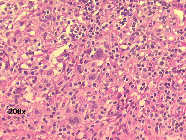
Hodgkin´s disease, FNA cervical lymph node, 4-year old girl


Drs. Prolla and Diehl's INTERESTING CASE OF THE MONTH January 2002 case A

FNA left cervical mass, large binucleated blast 500x M-G-G staining

FNA left cervical mass, Reed-Sternberg cell 500x M-G-G

FNA left cervical mass, Reed-Sternberg cell 500x M-G-G

FNA left cervical mass, Hodgkin's cell (left) - Reed-Sternberg cell (right) 500x M-G-G


Hodgkin´s lymphoma 200x H&E staining

Reed-Sternberg cell 500x H&E staining

Cells CD15 positive 500x

Cells CD30 positive 500x


The smears show Hodgkin's disease (HD) cellularity: several large binucleated cells with very large nucleoli (Reed-Sternberg cells), some large mononuclear cells with large nucleolus (Hodgkin's cells), and a background of lymphocytes and histiocytic/monocytic cells. Histology showed a mixed cellularity Hodgkin's disease, confirmed by CD15 and CD30 positivity, and CD45 and CD20 negativity in the immunohistochemical studies. Subtyping of HD by FNA material is not possible with specificity.
Note: Hodgkin lymphoma is suggested by the presence of Reed Sternberg cells in the setting of numerous lymphocytes on FNA. However, because this cell type is found in other conditions (eg, non-Hodgkin disease of the large-cell type), pathological identification of Hodgkin disease still requires additional tissue for surrounding cellular elements.


Features of Hodgkin´s disease (HD)- Romanowsky stains

Cellular patterns:
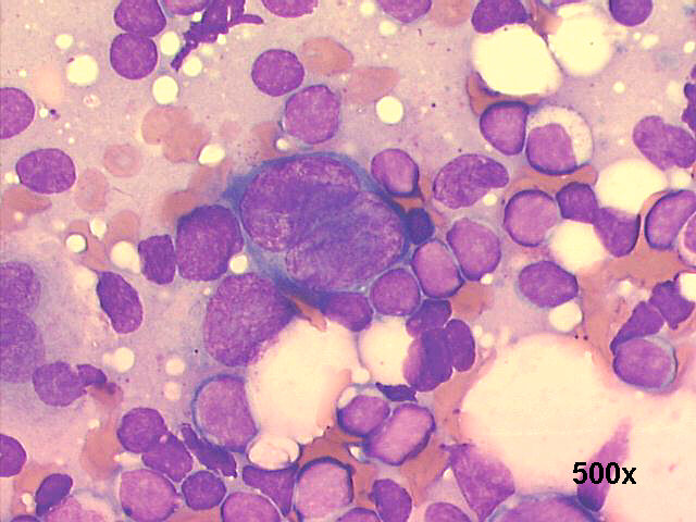
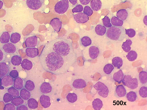
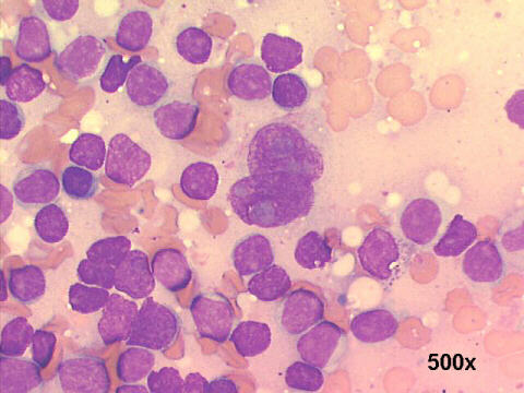
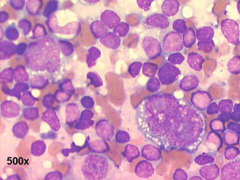
Polymorphic lymphoid cell population; "lymphoglandular bodies" in the background
Reed-Sternberg (R-S) cells: classic binucleated, with mirror-image nuclei. Hodgkin´s cells: mononuclear cell. Few multinucleated/polyploid cells.
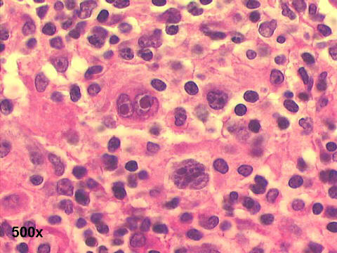
Cytoplasm:
Small amount of pale basophilic cytoplasm in R-S cells
Some cells have small cytoplasmic vacuoles
Nucleus
R-S cells: two large, hyperchromatic, mirror-image
R-S cells: each nucleus has a large macronucleolus. In Papanicolaou or H&E staining, with a clear halo around the nucleoli
Mitoses, karyorrhexis and apoptotic bodies may be found
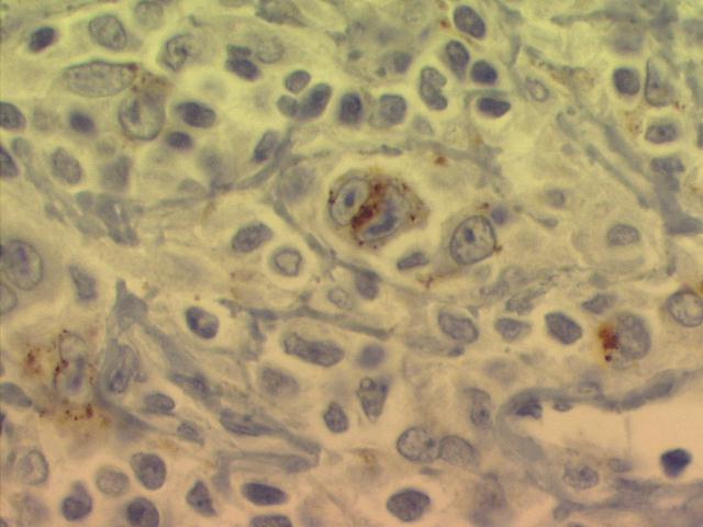
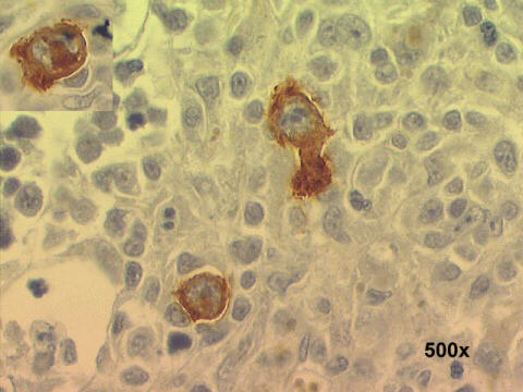
Phenotypic markers:
Classic R-S cells and its variants are CD15+ and CD30+, CD45-, CD20-
Lymphocyte-predominant HD is CD15-, CD30-, and CD45+
Caveat: FNA primary diagnosis of HD should be confirmed by surgical biopsy!


See also Case B


Back to Case 01A 2002 References
List of cases
Hosted by www.Geocities.ws

1 1