Drs. Prolla and Diehl's Case of the month ~ December 2006

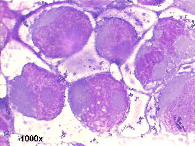
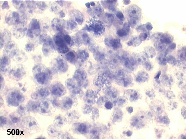
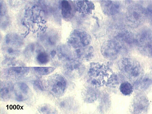
CNS fluid, 21-year old male, meningeal metastasis from testicular germ cell anaplastic tumor


testicular germ cell anaplastic tumor, 500x M-G-G staining, large anaplastic isolated cells

testicular germ cell anaplastic tumor, 1000x M-G-G staining, large anaplastic isolated cells, with huge nucleoli and scarce cytoplasm

testicular germ cell anaplastic tumor, 500x M-G-G staining, large anaplastic isolated cells

testicular germ cell anaplastic tumor, 1000x M-G-G staining, large anaplastic isolated cells, with huge nucleoli and scarce cytoplasm

testicular germ cell anaplastic tumor, 500x M-G-G staining, large anaplastic isolated cells

testicular germ cell anaplastic tumor, 500x M-G-G staining, large anaplastic isolated cells, with huge nucleoli and scarce cytoplasm

testicular germ cell anaplastic tumor, 1000x M-G-G staining, large anaplastic isolated cells, with huge nucleoli and scarce cytoplasm

testicular germ cell anaplastic tumor, 500x Pap staining, group of anaplastic cells, with huge nucleoli and scarce cytoplasm

testicular germ cell anaplastic tumor, 1000x Pap staining, group of anaplastic cells, two abnormal mitotic figures


Cytology smears revealed many large isolated anaplastic cells, compatible with metastatic lesion, primary germ cell tumor (embryonal carcinoma) in left testicle, previously treated.
The cytologic features of testicular germ cell tumors have been described by Balslev et al (Acta Cytol 34:690-694, 1990);
The present case is typical of the difficult differential morphological diagnosis between anaplastic carcinomas and anaplastic germ cell tumors. The immunecytochemistry may be very helpful: germ cell tumors being positive for PLAP (Placental-like alkaline phosphatase) and CD30, and negative for EMA (epithelial membrane antigen).
References:
Acta Cytol 41:497-503,1997


Case A December 2006 Case B December 2006 References List of cases Atlas
Hosted by www.Geocities.ws

1