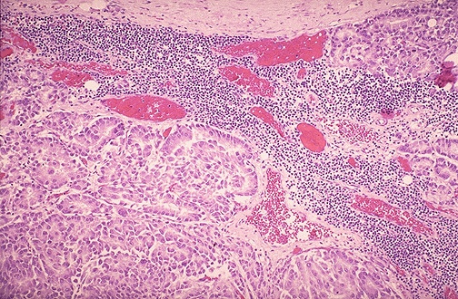
Lymph node: Metastases from SCC of lung
A
45 yr old Malay man complained of a swelling on the left side of his neck for 1
month. He
had coughed out fress blood several times last week.
Smoked 20 cigarettes a day since the age of 20.
Physical examination showed a painless, 2 cm diameter swelling at the
left side of his neck.
There were no chest signs.
Chest X-ray revealed a left lung shadow.
1.
What is the differential diagnosis?
Differentials include:
Primary lymph node disorders
Metastasis carcinoma
Reactive hyperplasia
Inflammation / infections
Lipid storage diseases
Endocrine diseases -
hyperthyroidism
2.
What
sort of biopsy would you recommend?
Bronchial
biopsy, fine needle aspiration of lung
Mediastinal
lymph nodes biopsy
3.
How
would you confirm a possible infective aetiology?
Ziehl-Neelsen
stain for acid fast bacilli � pink bacilli
4.
List
the histopathological features of the 2 conditions.
Tuberculosis:
Central
caseating necrosis surrounded by a rim of epitheloid macrophages followed by a
rim of lymphocytes and fibroblasts.
Langhan
giant cells may be present. (nuclei of these cells are arranged in a peripheral
horseshoe pattern)
Effacement
of lymph node architecture.
Metastases
from lung cancer:
Broad
sheets/ nests of squamous epithelial cells
Presence
of keratin pearls (lamellated pink stained masses)
Effacement
of lymph node architecture
5.
Name
a simple test to ascertain the nature of the lung lesion.
Microscopic examination of a bronchial biopsy and use Z-N stain for AFB.
<< PREVIOUS INDEX NEXT SLIDE >>
Copyright � Joseph Ong 2003