|
|
|
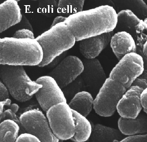
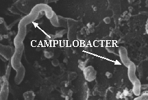
The size, shape and arrangement of bacteria, and other microbes, is the result of their genes and thus is a defining characteristic called MORPHOLOGY. Bacteria come in a bewildering and exciting variety of size and shapes, with new ones being discovered all the time. Nature loves VARIETY in its life forms as you can see just looking at your fellow humans. The most common bacterial shapes are RODS, COCCI, and SPIRAL. However, within each of these groups are hundreds of unique variations. Rods may be long, short, thick, thin, have rounded or pointed ends, thicker at one end than the other etc. Cocci may be large, small, or oval shaped to various degrees. Spiral shaped bacteria may be fat, thin, loose spirals or very tight spirals. The GROUP ASSOCIATIONS of microbes, both in liquid on solid medium, are also defining. Bacteria may exists mainly as single cells or as common grouping such as chains, uneven clusters, pairs, tetrads, octads and other packets. They may exist as masses embedded within a capsule. There are square bacteria, star-shaped bacteria, stalked bacteria, budding bacteria that grow in net-like arrangements and many other morphologies. When observing bacteria one should describe as many of these characteristics as possible. Consider how you would describe a BLIND DATE to a friend.
For an exceedingly colorful view of bacterial size and shape visit the Molecular Probe site. Click on the GALLERY and then on BACTERIA STAINS. Compare the size of Bacillus cereus and Pseudomonas aeruginosa. Click here for artificially colored bacteria.
![]()

Figure 3. Composite cartoon showing the major structures found in bacteria. No one bacteria contains all of these different components, but most bacteria contain the majority of them. Another GENERIC bacterial cell.
A typical bacterial cell is composed of the following structures. You are required to learn the function of each.
The CYTOPLASM is the TOTAL OF EVERYTHING INSIDE
of the cytoplasmic membrane. It has a gel-like consistency, but small
molecules can move through it rapidly; that is it only takes a few
microseconds for a molecule to move from one end of the cell to the other. The
following components are in the cytoplasm.
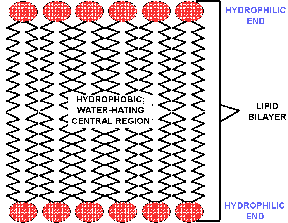
The CYTOPLASMIC MEMBRANE is the structure that
makes the cell possible as it is a SELECTIVE BARRIER that separates the highly organized
machinery of a cell from the frightful chaos on the exterior. The cytoplasmic
membrane is composed of a PHOSPHOLIPID
BILAYER in which are embedded the various proteins that control what goes
in and out of a cell (Views of membranes
and another
view that can be manipulated and still another
view that takes a lot of memory but shows much detail). The cytoplasmic
membrane is fluid (like a soap bubble) and usually very DELICATE and easily
ruptured if the supporting cell wall is removed. The inner portion of the
cytoplasmic membrane is composed of lipid or fat molecules that make it
impermeable to molecules that dissolve in water, but water itself can MOVE
FREELY through the cytoplasmic membrane. This inner region is said to be HYDROPHOBIC
or "water hating". Because the cytoplasmic membrane is a bilayer, the lipid
molecules face each other, placing the phosphate groups on the outer edges of
the cytoplasmic membrane. Phosphate is HYDROPHILIC
and likes to associate with water molecules. Many of the proteins embedded
in the lipid bilayer are TRANSPORT proteins that BIND specific molecules and
carry them into or out of the cell as required. The proteins allow the cell to
live in very dilute nutrient solutions because they soak up the nutrients like
a selective "sponge" and bring in the rare nutrient molecules. Click here
to look at some lipid molecules, chose the "gif" forms. | |
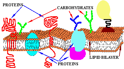
Figure 6. Cartoon of cytoplasmic membrane, showing its complex
composition of protein, carbohydrates and lipids. The proteins "float"
in the lipid "sea". Some proteins span the membrane, while others are attached
to only one side or the other, or to other proteins embedded in the cytoplasmic
membrane. Water can pass freely through the cytoplasmic membrane, but most other
molecules can not. One important characteristic of the cytoplasmic membrane is
that PROTONS (H+) are unable to cross the
cytoplasmic membrane. Click here
to learn about the lipid bilayer. Click here
to see more pictures of membranes along with a good discussion.
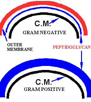
The CELL WALL is the total structure that defines the exterior of the cytoplasm. It includes the cytoplasmic membrane and one or two other layers in most prokaryotes. For a more detailed discussion of the bacterial cell wall composition click here. | |||
In GRAM NEGATIVE
cells the cell wall consists of three
components; the cytoplasmic membrane, a thin layer of a rigid sugar
MOLECULE NET immediately exterior to the cytoplasmic membrane that covers the
entire cytoplasmic membrane and an outermost lipid bilayer called the OUTER MEMBRANE. The rigid layer is called PEPTIDOGLYCAN and is the SHAPE-FORMING component of the cell.
The outer membrane also functions as a SELECTIVE GATE but its selectivity is much
LESS specific than that of the cytoplasmic membrane. The outer layer of the
outer membrane in Gram negative cells is composed of a molecule called
LPS. This molecule is medically
significant as it is HIGHLY TOXIC to humans and death from infections of G-
bacteria are often the result of poisoning by the LPS.
| Gram
positive cells have only two layers, the cytoplasmic membrane and a
THICK LAYER of
peptidoglycan as the outermost component. | |
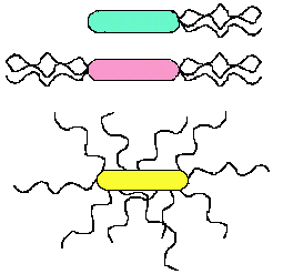
Several protein rod-like
structures pass through the cell wall. These include
the following:
|
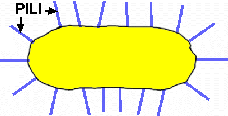
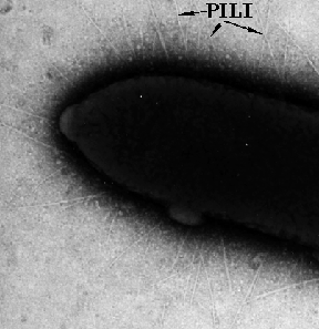
Figure 8. Pili. These fine hair-like, protein structures on the cell wall are pili. There are usually several 100/cell. In most cases they have special binding proteins at the end of the stiff rods. These types of pili are often important in adhesion of the cell to surfaces, such as teeth.
SEX
PILI are longer (than the adhesion-pili described
above), hollow protein rods that are mainly found on G- cells. These
structures are involved in the TRANSFER
OF GENETIC MATERIAL from one cell to another. The DNA that is transferred
may either be plasmid or chromosomal DNA. Cells which carry the genes for
making sex pili are said to be MALE or F+ cells. Usually the genes for sex
pili formation are carried by SEX
PLASMIDS. The process of DNA transfer is called CONJUGATION.
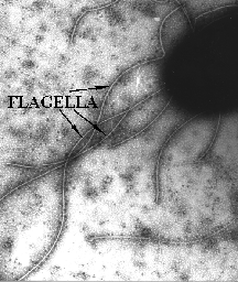
FLAGELLA are the final structures that
passes through the cell wall. Flagella are long,
rigid protein rods that provide movement to many motile bacteria. At their
base is a MOTOR that is driven by a
flow of PROTONS from the outside of the cell inward; much like a turbine in a dam is driven by
the flow of water through it. The number and arrangement of flagella on a cell
is part of its GENETIC CHARACTERISTICS and is used to describe each species. | |
The most exterior components of bacterial cells are the
CAPSULE and
SLIME layers.
These layers are usually composed of sugar polymers that are excreted by the
cell under certain conditions. The term capsule usually applies to a DEFINED
layer with a distinct outer edge, whereas a slime layer describes an
ILL DEFINED concentration of polymeric material which just slowly gets less
and less the further away from the cell. Although capsule production is a
genetic characteristic, its production is STRONGLY influenced by the nutrient
environment. For example, in a nutritionally poor medium a bacterium may
produce little or no capsule/slime, but in the presence of a high
concentration of sugar the capsule may be HUMONGOUS. The capsule has several
roles.
|
![]()
|
|
The ability of mobile cells to move in a desired direction is called TAXIS. Bacteria demonstrate several types of taxis. These include PHOTOTAXIS or the tendency to move TOWARDS or AWAY FROM LIGHT; CHEMOTAXIS. the ability to move TOWARDS a desired CHEMICAL or AWAY from a harmful one (POSITIVE or NEGATIVE taxis); MAGNETOTAXIS, the ability to follow the earth's MAGNETIC LINES OF FORCE. When you consider what these abilities REALLY MEAN you begin to achieve a true insight into the complexity of microbes. In the case of phototaxis the bacteria must have an EYE that detects light (i.e., they SEE light like you and I do). Furthermore different bacteria see different colored lights. In the case of chemotaxis, the bacteria have the equivalent of a NOSE in that they smell chemicals, identify them and then make a decision as to move away or towards them. That is, if they chemically detect a "nutrient" the motile ones move towards it, whereas if they "smell" a toxin, they move away from it. Magnetotactic bacteria have TINY MAGNETS in their cells that allow them to detect the north and south poles and then the ones in the northern hemisphere move north and those in the southern hemisphere move south. Which direction do you move in when you're hungry and smell pizza?
![]()
![]() CRITICAL THINKING QUESTION: How does the taxis of bacteria relate to
humans being bitten by mosquitoes?
CRITICAL THINKING QUESTION: How does the taxis of bacteria relate to
humans being bitten by mosquitoes?
![]()
|
|
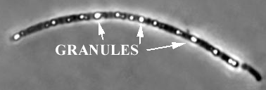
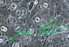
Some G+ bacteria form resistant structures called SPORES under adverse conditions. Spores are the most RESISTANT life form known. They are able to survive boiling in water at 100oC for long periods. Spores are resistant to UV-light, to drying and many harmful chemicals. We know spores can live for 100s of yr. and recently spores several million yr. old have been revived from insects trapped in amber. Some disease organisms like anthrax and botulism form spores that reside in the soil. The size, shape, and location of a spore in the cell are all identifying genetic characteristics. For example, in the figure below, the spore on the left is TERMINAL, OVAL and SMALLER than the cell.
![]()
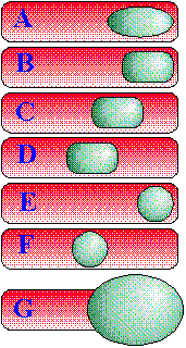
Figure 12. Spore structure and arrangements. The figure on the left shows the general structure of a bacterial ENDOSPORE. The figure on the right shows how the shape, location and the relative size of the formed-spore to the remains of the parent cell can be used to describe a bacterial spore-former. These characteristics are genetic and are like describing humans as being tall, blue eyed with blond hair. A = oval, terminal; B = rectangular, terminal; C = rectangular, subterminal, D = rectangular, central; E = circular, terminal; F = circular, central; G = terminal, club-shaped.
![]()
http://129.109.136.65/microbook/ch002.htm; Excellent chapter on bacterial structure and morphology.
![]()
Copyright © Dr. R. E. Hurlbert, 1999.
This material may be used for
educational purposes only and may not be duplicated for commercial purposes.