I. Classification of Sensory receptors
A. Mechanoreceptors: mechanical stimuli which responds to physical deformation, vibrational movement. ex: pressure applied to skin.
B. Chemoreceptors: amount or changing concentration of certain chemicals. ex: ion concentration, blood gases (O2, CO2)
C. Thermoreceptors: activated by heat or cold. sensitive to changes in temperature.
E. Nociceptors: activated by intense stimuli that results in tissue damage. results in pain
F. Photoreceptors: only found in eye. respond to light stimuli. detect and process light energy.
II. Somatic senses
A. Pain: Nociceptors are pain receptors widely distributed throughout the body. free nerve endings stimulated by tissue damage caused by mechanical stretching, chemicals, lack of blood flow, temperature extremes.
1. Types of nerve fibersa. acute fibers: used for sharp, intense, generally localized pain.b. chronic fibers: less intense, more persistent pain.
2. Types of pain
a. somatic pain: stimulation of superficial cutaneous receptors, deeper receptors in skeletal muscles, tendons, joints.b. visceral pain: stimulation of receptors in viscera.
B. Touch and Pressure
1. Meissner’s corpuscles, Krause’s end bulbs: smaller elongated receptors in dermal papillae in fingertips, palms, soles. touch, low frequency vibration, two-point discrimination.2. Ruffini’s corpuscles: small oval receptors made up of connective tissue capsule. deep pressure, sudden movement. located in dermis. slow adapting.
3. Pacinian corpuscles: large oval receptors of concentric layers of connective tissue around a central nerve ending. high frequency vibration, stretch. sensitive; adapt quickly.
C. Proprioceptors: located in muscles, tendons, joints, vestibular part of ear. awareness of position of body parts with respect to one another and movement of body parts.
1. Muscle spindles: complex sensory receptor in skeletal muscles. stimulated if length of relaxed muscle exceeds limit. result is stretch reflex. aids in maintenance of posture or positioning of body.2. Golgi tendon receptors: stimulated by excessive muscle contraction. sensitive to changes in tension on tendon. causes muscle to relax.
III. Special senses
A. Smell
1. Seven basic odors: camphorous, musky, floral, ethereal, pungent, putrid, pepperminty.2. Olfactory cell: modified bipolar neuron; dendrites end as coarse cilia: olfactory hairs. olfactory cilia extend from neurons in epithelial lining of upper surface of nasal cavity.
3. Olfactory mucosa: millions of olfactory cells, pigmented supporting cells, glandular cells.
4. Chemoreceptors are stimulated by gas molecules or chemicals dissolved in mucus. very sensitive, adapt rapidly.
B. Taste
1. Four basic tastesa. sweet (tip). sensitive to sugars, aldehydes, alcoholsb. sour (side). sensitive to acids.
c. bitter (back). sensitive to alkaloids, such as caffeine, nicotine.
d. salty (side). sensitive to ionized salts.
2. Taste receptors found on tongue.
3. Taste bud: taste cells and supporting epithelial cells.
a. circumvullate papillae (bitter)b. filiform papillae (sour)
c. fungiform papillae (salty)
4. Chemoreceptors stimulated by chemicals dissolved in saliva.
C.
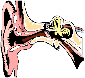
Hearing (auditory
apparatus) and Balance
(vestibular apparatus)
1. External ear: conducts and amplifies sound waves.a. Auricle (pinna): flap on side of head made of elastic cartilage.b. External Auditory Meatus: 2.5 cm tube leading into temporal bone. Cerumen (waxy product) produced here.
c. Tympanic Membrane: thin transparent membrane of fibrous connective tissue and epithelium. transmits sound waves to middle ear.
2. Middle ear: small, air-filled, epithelial-lined cavity in temporal bone.
a. Three ossicles:1. malleus: handle of hammer attached to inner surface of tympanic membrane.2. incus: head of hammer attaches to anvil by ligaments, moving as a unit.
3. stapes: anvil attaches to stirrup.
b. Eustachian tube: 4 cm auditory tube extending into pharynx. used to equalize pressure against inner and outer surfaces of tympanic membrane.
3. Inner ear: mechanoreceptors located here for the detection of stimuli for hearing and maintenance of equilibrium.
a. Bony labyrinth1. vestibule: oval window opens into it.2. cochlea: snail shaped structure in the anterior portion of the inner ear.
a. Organ of Corti: hearing sense organ found in cochlear duct.3. semicircular canals: superior, posterior, and lateral canals.
b. Membranous labyrinth: round window located at junction of vestibule and cochlea.
4. Sense of Hearing
a. sound waves enter external auditory canal.b. waves strike tympanic membrane, causing vibrations.
c. vibrations move malleus, then incus, then stapes.
d. stapes vibrates oval window
e. ripple in fluid in cochlea travels to fluid in cochlear duct to organ of Corti, eventually to round window.
f. movements of hair cells stimulates dendrites, initiates impulse to brain stem.
5. Equilibrium
a. Static Equilibrium: sense of position of head. located in vestibule, a bony chamber between semicircular canals and cochlea.b. Dynamic Equilibrium: motion of head. each semicircular canal contains membranous canal ending in ampulla. cristae ampullaris contain sensory hair cells which extend into the cupula, a dome-shaped gelatinous mass.
D. Sight
1. Visual Accessory Organsa. eyelids1. Function: cover and protect eyes.2. Structure
a. meiboman glands: large sebaceous glands to prevent tear overflow.b. orbicularis oculi (moves eyelid to close)
c. levator palpebrae superioris muscle (opens eye by raising upper lid).
b. conjunctiva: transparent layer of epithelium interspersed with goblet cells.
c. lacrimal gland: located in orbit, secretes tears continuously. series of ducts which carries tears to nasal cavity.
d. extrinsic eye muscles
1. superior rectus: rotates eye up, toward midline2. inferior rectus: rotates eye down, toward midline.
3. medial rectus: rotates eye toward midline.
4. lateral rectus: rotates eye away from midline.
5. superior oblique: rotates eye downward, away from midline.
6. inferior oblique: rotates eye upward, away from midline.
2. Structure of Eye
a. Outer tunic1. Cornea: transparent window that focuses light rays. covered anteriorly by nonkeratinizing stratified squamous, posteriorly by simple squamous.2. Sclera: white portion of eye. provides protection and attachment for external muscles.
b. Middle tunic
1. choroid coat: heavily pigmented elastic connective tissue. melanin used to absorb excess light; keeps inside of eye dark. prevents internal reflection.2. ciliary body: forms internal ring around front of eye. contains ciliary muscles. functions in adjustment for near and far vision.
3. lens: soft biconvex body enclosed in elastic capsule. held by suspensory ligaments. tension on ligaments changes shape of lens to allow for focusing.
4. iris: thin diaphragm of connective tissue and circular and radially arranged smooth muscle between cornea and lens. regulates size of pupil and amount of light entering the eye.
c. Inner tunic
1. retina: inner lining of eyeball which contains photoreceptors.2. fovea capitis: depression in retina where sharpest vision is produced.
3. optic disk: where nerve fibers and blood vessels leave eye: the blind spot.
4. posterior cavity: filled with vitreous humor, which supports the eye and maintains its shape.
3. Visual receptors: modified neurons. photoreceptor cells that detect and process light.
a. rods: long thin projections located at terminal ends. more light sensitive, allow vision in dim light.b. cones: short, blunt projections. allow for color vision, sharp images.
4. Visual pigments
a. Rods: rhodopsin (visual purple): breaks down into opsin and retinal.b. Cones: retinal combines with one of three proteins.
1. erythrolabe: detects red light waves.2. chlorolabe: detects green light waves.
3. cyanolabe: detects blue light waves.
5. Physiology of Vision
a. Four processes1. Refraction of light: light rays bend when traveling from one medium to another. through eye: four different media: cornea, aqueous humor, lens, vitreous humor.2. Accommodation of lens: convex lens: thickest at center. converge light rays to focal point. image formed on retina inverted and reversed.
3. Constriction of pupil: regulates amount of light to minimize scattering, ensure optimal exposure of light to retina.
4. Convergence of eyes: for binocular vision. coordinated actions of extrinsic eye muscles move eyes slightly medially so visual axes converge.
b. Emmetropia: normal vision.
c. Myopia: nearsighted.
d. Hyperopia: farsightedness.
e. Presbyopia: loss of elasticity of lens with age.
f. Astigmatism: curvature and thickness of cornea or lens not uniform.