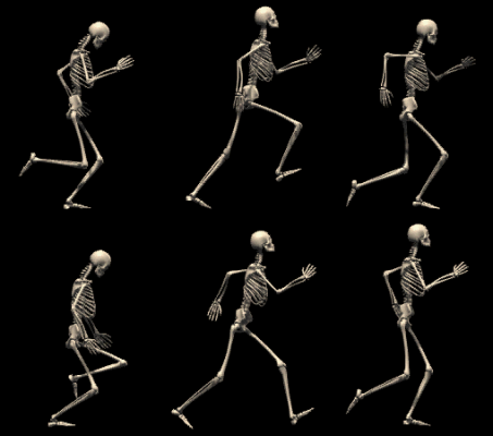

I. Functions of bone
A. Support: supporting framework . shape, alignment, positioning
B. Protection: delicate structures underneath
C. Movement: act as levers moved by contraction of muscles.
D. Mineral storage: Ca, P, etc. helps in homeostasis.
E. Hematopoiesis: blood cell formation: in ends of long bones, flat bones of skull, pelvis, sternum, ribs.
II. Types of Bones
A. Long bones
1. diaphyses: main shaft-like portion. thick compact bone, hollow cylindrical shape. functions to provide strong support, less weight.2. epiphyses: ends of long bone. bulbous shape for joint stability, attachment of muscles. spongy bone located here, filled with red marrow. separated from diaphyses by epiphyseal plate.
3. articular cartilage: thin layer of hyaline cartilage. covers joint surfaces. cushions jars and blows.
4. periosteum: covering of bone made of dense white fibrous tissue. fibers penetrate bone to weld together. muscle tendon fibers interlace to attachment to bones. contains small blood vessels that branch into bone. essential for bone cell survival, bone formation.
5. medullary cavity: hollow space in diaphysis. filled with yellow marrow rich in fat.
6. endosteum: thin epithelial membrane lining medullary cavity
B. Short, Flat Irregular bones have inner portion of spongy bone, covered with compact bone.
III. Bone Tissue
A. Two types of bone tissue
1. Compact bone: dense, solid2. Spongy bone: open space filled by assemblage of needlelike structures
B. Matrix composition
1. Inorganic salts: deposits of chemical crystals of calcium and phosphate. Mg, Na also found.2. Organic matrix: ground substance of protein, polysaccharides surrounds collagenous fibers. gives strength, plastic-like resilience.
C. Microscopic structure of compact bone: cylindrical osteon (Haversian systems) surrounding canals running lengthwise through bones. permits delivery of nutrient, removal of waste products.
1. lamellae: concentric cylindrical layers of calcified matrix2. lacunae: small spaces containing tissue fluid in which bone cells lie.
3. canaliculi: ultra small canals radiating from lacunae connecting them to each other and to Haversian canal.
4. Haversian canal: extends lengthwise through center of osteon. contains blood vessels, lymphatic vessels, nerves.
5. transverse canals (Volkman's) connect osteons.
D. Microscopic structure of spongy bone: no osteons.
1. trabeculae: needlelike bony spicules.2. canaliculi transport nutrients, wastes to and from cells.
3. spicules arranged along lines of stress to enhance bone strength.
E. Bone Cells
1. Osteoblasts: bone-forming cells. synthesize and secrete osteoid (organic matrix) that is deposited in framework of collagen fibrils. results in accumulation of mineralized bone.2. Osteoclasts: giant multinucleate bone-reabsorbing cells. responsible for erosion of bone minerals. activity alternates between osteoblasts and osteoclasts.
3. Osteocytes: mature, non-dividing osteoblasts surrounded by matrix. cytoplasmic extensions extend into canaliculi.
F. Bone Marrow
1. myeloid tissue: soft, diffuse connective tissue.2. Function: site of blood cell production.
3. Location: found in medullary bone cavity of long bone, spaces of spongy bone. in child, nearly all marrow is red marrow. replaced by yellow marrow as one ages. saturated with fat, therefore no blood cell production.
IV. Development of bone
A. Intramembranous ossification: occurs in some flat bones (skull).
1. centers of ossification: groups of osteoblasts produce organic matrix.2. trabeculae appear, joint to form network: spongy bone.
3. flat bones grow by adding osseous tissue to its outer surface.
B. Endochondral Ossification (occurs in long bones)
1. cartilage model develops periosteum
2. collar of bone deposited by osteoblasts formed by enlarging of periosteum.
3. cartilage begins to calcify.
4. blood vessel enters at midpoint of diaphyses, forms primary ossification center. ossification progresses toward epiphyses, bone grows in length.
5. secondary ossification centers appear in epiphyses.
C. Epiphyseal plate: cartilage plate joining epiphysis and diaphysis (growth plate). bone is finished growing when epiphyseal plate completely calcified.
V. Healing a Fracture
A. Fracture hematoma: after break, blood clot forms.
B. Callus forms as bloodclot is reabsorbed. binds broken ends together inside and out. stabilizes fracture.
C. Callus modeled, replaced with normal bone.
D. Injury healed completely.
VI. Cartilage
A. Hyaline: most abundant. appears like milk glass.
1. covers articular surfaces of bone.2. forms costal cartilages to connect ribs to sternum
3. forms cartilage rings to connect ribs to sternum.
4. tip of nose
B. Elastic cartilage: contains some collagen, more elastic fibers.
1. external ear2. epiglottis
3. eustachian tubes
C. Fibrocartilage: small amount of matrix, abundant fibrous element.
1. symphysis pubis2. intervertebral disks
3. near points of attachment of large tendons.