PATHOLOGY
Patho (disease, misery) and logy (study of, reasoning about).
Pathology is study of disease.
More appropriately is study of morphological, functional or biochemical changes in cells, tissues or fluids of the body.
From the ancient times till now there has been a mammoth increase in the knowledge about disease which has gone from macro to micro level.
Pathology is study of appreciating something wrong in the body which for the student is what,
further questions then arise that why and how this wrong has happened or developed.
The subject of GENERAL PATHOLOGY (a branch of pathology) revolves around the mechanisms by which tissues are injured together with the expressions of injury and the functional consequences of it,
BRANCHES OF PATHOLOGY
Medical Pathology deals with study of disease in man,
Veterinary Pathology deals with study of disease in animals.
Diagnostic Pathology deals with identification of nature of disease using traditionally, gross or histopathological (microscopic pathology) tools but now a days other tools are also used in the subject.
Forensic Pathology deals with medicolegal issues, e.g., ruling of death of animals by poisoning in which case court or police department is involved.
Surgical Pathology deals with study of biopsy material (tissue taken during operation).
Environmetal Pathology deals with study of effects of environment in the causation of disease or impact of environment on disease
Toxicological pathology deals with study of effects of different toxic substances in the causation of disease.
Zoo and Wild life Pathology deals with diseases in zoo and wild animals, respectively.
Immunopathology is a branch of pathology that deals with tissue injury produced by the involvement of immune system of the body.
Clinical Pathology deals with laboratory analysis of disease including haematology, cytology, serum chemistry, endocrinology, urinalysis etc.
Experimental Pathology deals with experimental study of diseases that may involve experimental production of disease and then study of different abnormalities produced thereto.
General pathology is the branch of pathology that deals with common denominators (lesions) of disease and mechanism of production of these.
Systemic pathology is a branch of pathology that deals with study of diseases with reference to system of involvement and is sometimes also named special pathology.
LANGUAGE OF PATHOLOGY
Lesion is a term used to denote an abnormality and mostly sound for any structural abnormality but rarely is used to describe the functional abnormality or biochemical lesion. The examples includes abscess, extra growth, change or missing enzyme or biochemical etc.
Pathognomonic is a term which means that a particular abnormality is found only in specific condition, e.g., hearing the fetal heart tone is pathognomonic of pregnancy.
A biopsy is tissue or peace of tissue removed from an animal during life for diagnostic purpose. This is mostly obtained to make clear a suspected cancerous from non-cancerous outgrowths
The autopsy ("necropsy") is the opposite of "biopsy". It means a pathologist examines part or all of a dead body by doing postmortem. The term autopsy is used in human discipline, while necropsy is used in veterinary discipline with same meaning.
The Disease can simply be described by breaking the word into two, i.e., dis and ease which is self explanatory. However, disease is term used to describe clinically apparent dysfunction due to abnormalities, excesses, deficiencies, and injuries occurring at the cell and tissue level.
Pathogenesis is a term used to describe the mechanism of development of disease and involves step-by-step progression of disease.
Diagnosis means naming a disease on the basis of clinical signs, laboratory test and lesions characteristics of disease , e.g., pericarditis and perihepatitis (lesions) in poultry are very much characteristic of colibacillosis (disease).
Prognosis is a term used to designate the possible outcome of disease whether favourable or unfavourable or in other words good or poor.
Aetiology refers to cause of disease that may be due to intrinsic or extrinsic reasons, e.g., tuberculosis is caused by Mycobacterium (bacteria).
Clinical signs and symptoms: signs are evidence of disease discovered by the physician and are used to describe abnormal behaviour by animals as exhibited by them due to disease, e.g., higher temperature, staggering gait, yellow discolouration of eyes and mucus membranes.
A syndrome is a cluster of symptoms and/or signs not necessarily due to the same cause. e.g., malabsorption syndrome.
TOOLS OF PATHOLOGY
- Eyes and Hands
- Light Microscope
- Histochemical Stains — Fat - Oil red O, Best Carmine - Glycogen, Acid Fast - Mycobacterium etc.
- Darkfield, Phase Contrast, Fluorescence microscopy
- Electron Microscopy
- Transmission - thin Sections
- Scanning - Three dimensional structure
- Photometers / Colourimeters etc.
NORMAL ---- ADAPTATION --- INJURY
NORMAL – HOMEOSTASIS
Maintenance of physicochemical activity compatible with cell survival and functions.
ADAPTATION
Establishment of new level of physicochemical activity still preserving health of itself.
INJURY
Loss of ability of cell to maintain normal of adaptive homeostatic state.
ADAPTATION
PRINCIPAL
Atrophy
Hypertrophy
Hyperplasia
Metaplasia
ATROPHY
Organ Atrophy — Cell Atrophy ?
Causes
reduction in work load
loss of innervation,
loss of endocrine stimulation,
reduced blood supply or inadequate nutrition,
pressure
aging
The cellular change is identical in all cells
Example — Leg muscles of fixed or immobilized leg in fracture.
The biochemical mechanism of atrophy is poorly understood, in particular the signal that triggers the process.
Atrophy may be numerical (number) or quantitative (cell protoplasm)
Catabolic processes exceed anabolic processes – autophagic vacuoles
There may be "residual bodies"
Microscopically
Cells may be fewer (numerical) or smaller than normal (quantitative atrophy).
In spleen, the trabeculae – large or too numerous
In kidney, the glomeruli – too numerous and closely associated
In atrophic muscles, – sarcoplasm narrower and disappears – leaving sarcolemma.
In liver, the hepatic cords – become thinner.
Missing cells replaced by adipose tissue as in physiologic atrophy of the thymus Fatty atrophy:
Sometimes replaced by fibrous connective tissue Fibrous atrophy:
Grossly
The organ or part is smaller than normal.
It is well appreciated in paired organs where one organ is reduced in size and weight compared with its counterpart.
HYPERTROPHY
- Increase in size of cells and thus increase in size of organ or tissue
- Occur in tissue whose cells can not divide – STRIATED MUSCLES
- Organ Hypertrophy – Cellular Hypertrophy (Increase in size)
- Increase in size of most tissues and organs — hypertrophy and hyperplasia
- Pure hypertrophy occurs in tissues whose cells cannot divide.
- The nature of the signal to the cell is poorly understood.
- There is an increase in total cellular proteins, including myofibril in muscle cells and organelles such as mitochondria, ER and myofilaments.
- The anabolic processes exceed catabolic processes.
- The metabolic activity per unit volume — close to normal cell
- There may be hypertrophy of specific organelle system, e.g., SER.
PHYSIOLOGICAL
Skeletal muscles ---- weight lifters, athletes Hormonal stimulation — Uterus smooth muscles during pregnancy – oestrogen PATHOLOGICAL
- Myocardial hypertrophy — stenotic (narrowing) valvular diseases or lung diseases.
Microscopically
- The cells are larger than normal.
- Cells can be compared with those of other organ from the same animal or from other animal of same age, whatsoever is suitable.
Grossly
- The organ/tissue appear enlarged
- Can be compared with animal of same age and breed.
- Seen in organs have no potential of mitotic activity
HYPERPLASIA
It is the increase in size of an organ due to increase in number of its constituent cells
Occur in those organs/tissues whose cells can divide
Hyperplasia after the removal of the stimuli does not progresses.
Hyperplasia of connective tissue and blood vessels (together granulation tissue) in wound healing is an important response.
PHYSIOLOGICAL
Increased level of a normal stimulus (hormonal),
In regeneration – liver regeneration after injury.
As a compensatory response, e.g.,
missing organ in paired organ,
partial resection
PATHOLOGICAL
Pathological hyperplasia, occur because of excessive response or due to excessive stimulation.
Hyperplasia of thyroid gland (goiter) — iodine deficiency
Hyperplasia of bronchial epithelium — chronic irritation
Hormonal Stimulation – uterine – abnormally high oestrogen
Excessive growth factor stimulation — warts – papillomaviruses
Microscopically
The cells are more numerous than normal number.
Some cells may have mitotic activity (can confuse with neoplasia)
Cell are highly differentiated
Cells can be compared with those of other organ from the same animal or from other animal of same age, whatsoever is suitable.
Grossly
The organ/tissue appear enlarged
Can be compared with animal of same age and breed.
Seen in organs/tissue have potential of mitosis
METAPLASIA
Mature differentiated (highly specialized) cell type is replaced by another differentiated cell (less specialized ) type.
Change occurs as a response to better withstand the adverse environment
The change is thought to occur due to genetic reprogramming of the epithelial stem cells or of undifferentiated mesenchymal cells.
Occurs mostly due to chronic irritation, including inflammation.
Change of columnar epithelium of trachea, bronchi and bronchioles into stratified squamous epithelium in cigarette smokers is an example of metaplasia.
Vitamin-A deficiency results in wide spread squamous metaplasia in the body.
In most of the cases, metaplasia depends on proliferation of germinal cells or stem cells whose progeny undergo modified differentiation.
In fewer cases, direct transformation of differentiated cells takes place.
Microscopically
In mesenchymal metaplasia, the fibrous tissue may get replaced by bone or cartilage.
Osseous metaplasia occurs as an ageing process in cartilage, e.g., the costal and thyroid cartilages.
Osseous metaplasia is also seen in soft tissues, as in scars and in areas of dystrophic calcification.
There is change in differentiated cell type from that present in routine in a tissue.
Grossly
Difficult to appreciate grossly
DYSPLASIA
Is abnormal development of tissue or disorderly growth, or malformation of tissue.
The term is more often used to describe a proliferative response (non-neoplastic) accompanied by loss of regular differentiation.
The change is considered as preneoplastic.
Microscopically
There is cell atypia and disorderliness.
It is loss of uniformity of the individual cells and loss of architectural orientation.
Cellular atypia is characterized by pleomorphism and hyperchromasia
Mitotic figures are seen in abundant cells.
Grossly
No much can be appreciated grossly
APLASIA
The term refers to failure to grow of an organ and there may be presence of a rudimentary organ.
This is abnormality in growth which may be genetic.
Haematologist use the term where the bone marrow undergoes destruction
And there is no regeneration and no new cell formation
Grossly
Absence of or rudimentary organ
HYPOPLASIA
The term is used to refer to failure of an organ to reach its normal full adult size.
It is arrested growth.
Examples include renal hypoplasia and hypoplasia of the testis.
The causes include, (1) genetic mutations (hereditary), and (2) deletion of critical cell population during development, may be in utero, due to certain viruses, toxins and irradiation.
Congenital cerebellar hypoplasia in young animals can occur due to viral infection during pregnancy.
Hypoplasia of kidney, pituitary and pancreas can occur spontaneously and is hereditary.
Microscopically
Fewer cells than normal
Size of cells may also be small
Grossly
Smaller than normal organ
NEOPLASIA
The term is used to refer to new growth, i.e., an excessive and persistent tissue growth which is not under the growth control mechanism of the body
The growth continues even after the cessation or removal of the stimulus.
Both benign and malignant tumors are clonal overgrowths of cells bearing multiple genetic injuries.
Tumor cells typically have failure of division control, failure of senescence ("immortalization"), and failure of proper apoptosis.
Cancer cells can be characterized as antisocial, fairly autonomous units that appear to be indifferent to the constraints and regulatory signals imposed on normal cells.
For further detail, the reader is referred to chapter on neoplasia.
ATRESIA
It is the complete failure of the lumen, or a portion of the length of the lumen to form
The condition is congenital and the animal is born with the defect.
The most common occurrence is atresia-ani, i.e., there is no anal opening right from the birth.
It is a non-neoplastic narrowing of a lumen.
It may be a birth defect, or acquired.
Occlusion is the complete obstruction of lumen that was once open.
Atherosclerosis (narrowing of lumen of blood vessels), the disease results when phagocytic cells in the intimal layers of large arteries become engorged with cholesterol and its esters.
The cholesterol (esters) in tissue sections can be recognized by the "needle-shaped" clear spaces left behind as it is removed in processing ( Cholesterol crystals are really flat rectangles in cross section).
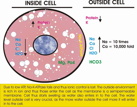
CELL INJURY
Cell Injury: Mechanism of cell injury by interfering with energy production, Hypoxic injury
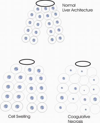
SCHEMATIC DIAGRAM OF CELL SWELLING AND COAGULATIVE NECROSIS -LIVER
CAUSES OF CELL INJURY
HYPOXIA
(lack of oxygen),Ischaemia
Occlusion of the blood vessels
Failure of the heart to pump enough blood
Hypoxaemia:
Too little oxygen in the blood
Too little oxygen in the air
Failure to properly ventilate the lungs
Failure of the lungs to properly oxygenate the blood
Failure of the heart to pump enough blood through the lungs
Tremendously increased dead space (i.e., pulmonary thromboembolus)
Haemoglobin problems ("anaemic hypoxia")
Inadequate circulating red cell mass ("anaemia")
Inability of haemoglobin to carry the oxygen (carbon monoxide poisoning, methaemoglobinemia)
High affinity haemoglobins that will not give up their oxygen to the tissues
Failure of the cytochromes ("histotoxic hypoxia")
Cyanide poisoning
Rotenone poisoning
Dinitrophenol poisoning
LACK OF NERVE SUPPLY
Horses used unprepared for heavy work load
CHEMICALS AND DRUGS
Direct reacting Cyanide, heavy metals
Indirect reacting Carbon tetrachloride
Acids and alkalies
PHYSICAL AGENTS
Mechanical injury (crush injury, fractures, lacerations, haemorrhage)
Extremes of heat or cold
Radiation - (x-rays, radioactive elements, ultraviolet radiation)
Electric shock
Sudden changes in atmospheric pressure (blast injury, decompression injury in divers)
Noise trauma.
GENETIC DEFECTS
Congenital malformations.
Genetic factors can produce diseases directly or influence its outcome.
Inherited diseases
Patent ductus arteriosus (dog),
lysosomal storage disease,
combined immunodeficiency (horse)
Genetic influence on susceptibility to disease
Canine distemper, a disease of young pups
BIOLOGIC AGENTS
Bacteria, Viruses, Prion, Mycoplasma, Chlamydia etc.
NUTRIONAL IMBALANCES
Deficiencies Se, Vitamin E, Protein, Carbohydrate etc.
Excesses Fat – cholesterol
IMMUNE MECHANISM
Allergic anaphylactic reactions mediated by IgE antibodies
Cytotoxic reactions during which complement is activated
- Autoimmune reactions