Rhinology/Allergy pages
Cerebrospinal fluid fistula repair
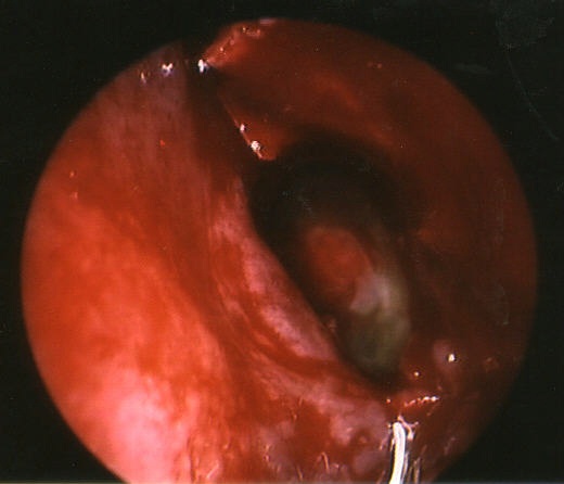
Left sphenoidotomy with pooling of green-colored fluid. The fluid is fluoroscein-stained cerebrospinal fluid (csf).
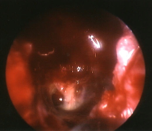
The same left sphenoid with the scope advanced. The hole itself can just be seen. It is the blood-tinged hole in the lower left side of the picture; pale mucosa is seen next to the hole.
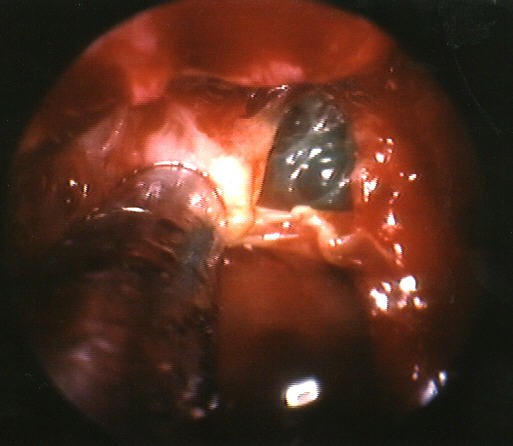
Another patient with a left sphenoid sinus CSF leak that had resulted in several bouts of severe meningitis. The face of the sphenoid has been taken down and we are looking into the sinus itself. The suction is pulling on the mucosal flap that partially covered the superior sphenoid sinus.
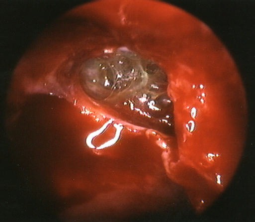
A closer view of the patient above. The delicate fibrous tissue is either pia mater or necrosed brain tissue. the yellow-staining is due to fluoroscein dye. This patient suffered their csf leak as a result of a trauma several years earlier. He has been "meningitis-free" since surgery.