| Sinus CT evaluation |
 |
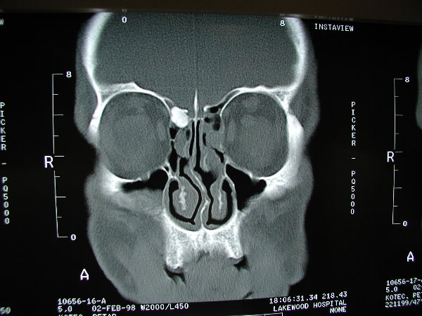 |
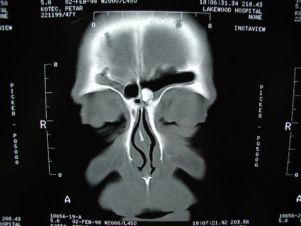
| The patient above had two separate osteomas within his sinuses. On the right it is within the frontal sinus outflow tract, and on the left it is within the frontal sinus iteslf. The osteoma on the right was very symptomatic, causing obstruction and severe pain, while the left was asymptomatic. The right was removed endoscopically, and the left is being observed for the time being. An osteoma is a benign boney growth that can grow and occlude paranasal sinuses and cause problems. |
 |
 |
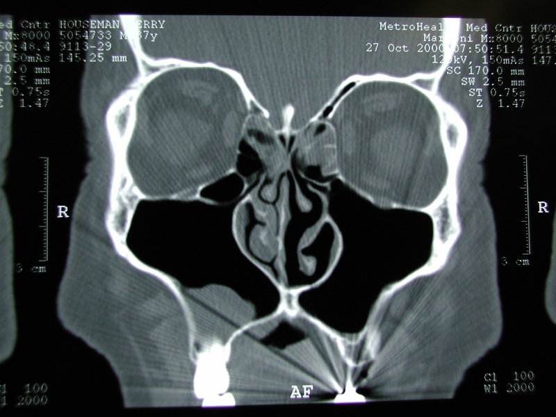 |
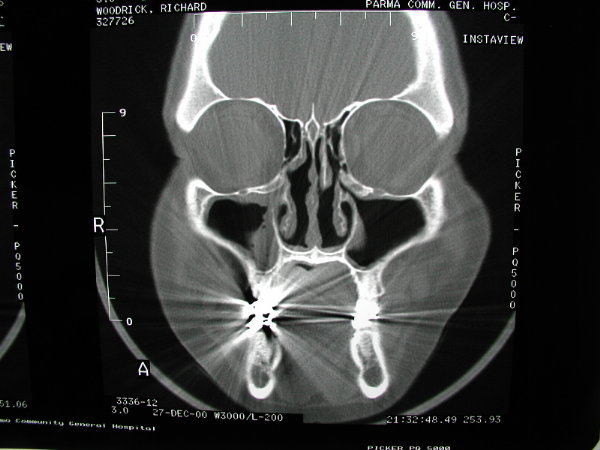
| The patient above had previous sinus surgery. Maxillary antrostomies were created which did not incorporate the patient's natural maxillary antrostomies, hence his symptoms persisted. Careful evaluation of his CTs and appropriate connection of his natural to iatrogenic ostia led to recovery. |
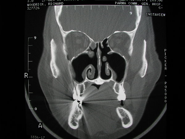
| The patient above had a fascinating CT scan. He basically has no nasal floor, as the maxillary sinuses extend below his nasal cavity, and replace his hard palate. His sinus surgery was surprisingly straight-forward and he healed rapidly. |