| CRANIOSYNOSTOSIS |
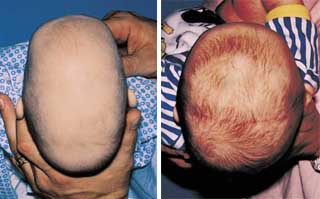
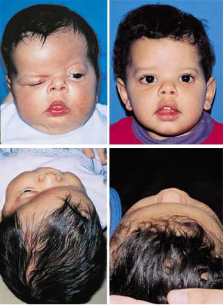
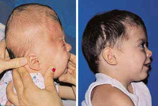
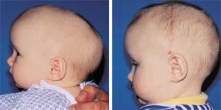
| Craniosynostosis is known as the premature closing of one or more of the bony gaps in an infants skull. These gaps are known as the cranial sutures. In a normal infant not affected with craniosynostosis these sutures remain open throughout the first two year of life to allow for proper brain growth. The skull shape is affected by craniosynostosis according to which suture closes. Craniosynostosis is known to happen in about one in every 1500 births. The only treatment for Craniosynstosis is surgery. However, as further research is done more surgical options are becoming available. Intercranial pressure and learning delays can be a few of the complications associated with uncorrected Craniosynostosis. A few other concerns are eyesight problems, hearing delays, speech and mouth problems, and hydrocephalous(an abnormal collection of fluid on the brain which can lead to pressure if left untreated), to name a few. If Craniosynostosis is suspected the advise of a specialist should be sought out and a CAT Scan should be performed. An X-Ray can not rule out Craniosynostosis and should always be followed with A CAT Scan. |
| Terminology of Craniosynostosis |
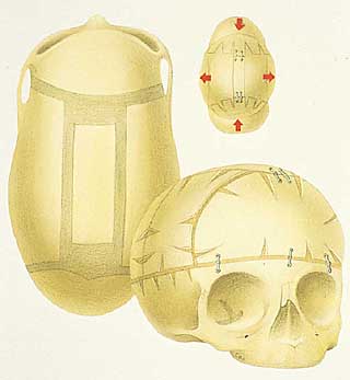
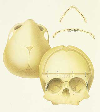
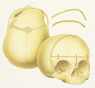
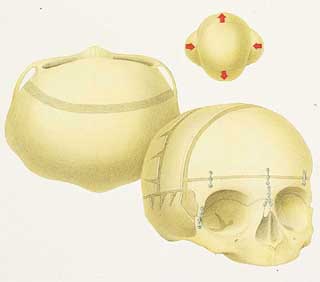
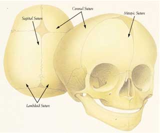
| FUSED SUTURE ______________ Sagittal ______________ Metopic ______________ Unilateral Coronal ______________ Bicoronal ______________ Lambdoid |
| NAME ______________ Scaphocephaly ______________ Trigonocephaly ______________ Plagiocephaly ______________ Brachycephaly ______________ Plagiocephaly |
| DESCRIPTION _______________ Boat Skull _______________ Triangular Skull _______________ Asymmetric Skull _______________ Short Skull _______________ Asymmetric Skull |
| Click on Picture for digram of Sutures |
 |
| History of Craniosynostosis and Surgery |
| In 1830 the term craniosynostosis was coined by a Dr. Otto. In 1851 Dr. Stahl and Dr. Hyrtl noticed that the premature closure of the cranial vault sutures led to abnormal head shape and in 1851 Dr. Virchow described how the skull will grow parallel to the affected suture. Dr. Virchow is also the first person to classify the deformity according to the sutures involved. It was only in 1910 to the 1920s that observations of more complex cranial and facial deformities led to the syndromes known as Crouzon(abnormal growth of the facial skeleton) and Aperts(abnormal growth of facial skeleton along with limb deformities.) Dr. Moss discovered in the 1950's that removal of the affected suture may not alter the abnormal skull growth in sagittal and bicoronal patients. He concluded that the main cause of the cranial deformity in mosty types of craniosynostosis starts at the skull base rather than at the fused sutures. These views led to change in surgical treatment of craniosynstosis. No longer were affected sutures just removed but complex cranial expansion procedures and remodeling were put into practice. Dr. Tessier was the first to attempt major surgical procedures on the craniofacial skeleton initially performing the procedures as part of reconstructive surgery following trauma and later attempted on patients with craniosynstosis. Dr. Tessier pioneered the craniovault reconstruction including fronto-orbital and midface advancements, he is known as the father of modern craniofacial surgery. Introduction of the CT scan in the 1970's led to new understanding and visualizations of the effect of craniosynostosis on skull growth thus added to the way traditional craniovault surgery is performed to this day.. Craniosynostosis is estimated to happen one case in every 1500 live births with case of Couzon syndrome 1/60,000, Aperts 1/10,000, Saethre-Chotzen and Pfeiffer syndromes being the rarest. Clinical problems associated with craniosynostosis if left untreated can be airway problems, sleep apnea, speech and growth delays, chronic headaches, visual failuers and negative social effect due to abnormal facial/head appearance. Surgery appears to be more successful when done before 9 months of age with less risk of reoperation. Increasing awareness of craniosynostosis keeps surgical options open to children if the condition is caught early enough. Don't be afraid to push to see a specialist if you suspect something is wrong your child. |
| Click below for example of head shapes |
| Leigha's Information Sheet PRINT ONE OUT This is a Guide to help reconize the warning signs of Craniosynostosis. It is a simple 2 page overview that should be shared with EVERYONE. Please print these out and share with friends, doctors, anyone you feel would be interrested. Thankyou! |