|
| Costel Rizescu |
|
R & D Activity / Paper Abstracts |
![]()
APPLICATIONS IN: A. GEOLOGY AND MARINE RESURCES B. ARCHAEOLOGICAL & HISTORICAL SCIENCE |
|
|
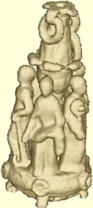
Neutralisator - work of art made by gold (belonging to Romanian Art Museum). Photo and 3-D tomogram Click here to see 3-D animation |
I worked in manufacturing of first CT systems in Romania, developing and implementing applications for end-users. I have been responsible for all aspects, from design, manufacturing, and installation through operation, testing, and field use of CT systems with gamma-ray sources (137-Cs, 192-Ir) and, later, with X-ray tubes. Early in my career I focussed on using the 192-Ir radioisotope in the CT scanner in order to employ a newly developed dual-energy method. It better characterizes the interior structure by mapping local density and effective atomic number, thus extending the capability of quantitative analysis.
Between 1993 and 1997, I authored the dual-energy method, developed and applied it with significant results on industrial objects as composite and sintered materials, ceramics, and materials for defense industry. Subsequently, I applied the same method in other fields of science.
In geology and paleontology, I determined structural parameters of sedimentary and metamorphic rocks, polymetallic nodules, corals, fossils, and marine cores, such as: degree of mineralization, total or partial porosity, volumetric and mass concentration of fractions of material, bulk/local density and effective atomic number, and stratigraphy frequency. In historical science and archaeology, I scanned hundreds of artifacts, I analyzed and detected counterfeit works of art, identified restoration and preservation procedures for artifacts presenting severe deterioration, and determined internal components of works of art for evaluation purposes. In forestry research, I pursued technical solutions for mobile CT scanners and applied them in determining the state of health of trees, ages, wood density and humidity. Also, I proposed an environmental research method for identifying the weather conditions history by scanning wood artifacts, and I have been involved in developing procedures for identifying drugs and illegal weapons transiting airports and customs check points.
I consider the X-ray dual energy CT method can offer precious information on structural composition of samples with low-density and low-Z phases by using high detection resolution systems and maneuverability of ranging a large X-ray spectrum. After defending my Ph.D. thesis in 2001, I intended to conduct a follow-up study on parametrizing the dual-energy CT method for energies in the range 70 - 1500 keV, thus extending its applications area in investigating radioactive waste. Since in the last years my career changed towards the medical field, this and other scientific goals have not materialized yet; however, presently I would be very interested to pursue them and thus resume my research activity in applied physics.
Paper Abstract This is a newly CT method in determining local density and effective atomic number using dual-energy gamma radiation from 192-Ir radioisotope (Rizescu, C., Beşliu, C., Jipa, A. (2001) Determination of local density and effective atomic number by the dual-energy computerized tomography method with 192Ir radioisotope, Nuclear Instruments and Methods in Physics Research I, A 465, 584 - 599).
The applied method is based on: 1) modeling total effective cross-section of gamma radiation in the natural materials in function of the atomic number (Z from 1 to 92), in terms of the photoelectric and Compton cross-sections, and 2) using a new parameter (composition factor k) given by the ratio of the linear attenuation coefficients from the two tomograms.
The authors investigated 16 materials with effective atomic number Zeff between 6 and 72.2 and bulk density ranging from 2.15 g/cm3 to 10.51 g/cm3, using 310.5 keV and 469.1 keV gamma radiation. The results show very good accuracy of the dual-energy CT method for most materials. The error in determining the effective atomic number is less than 5% for Zeff >25 and less than 10% for light materials (Zeff <15), while the error in density estimation is always smaller than 3%. go to top

Original collimator for micro-tomography
Sketch of micro-collimator for X and γ-rays computer tomographs
2-D tomogram with Hg micro-collimator.
Material: cooper wires; Source: 192-iridium, Energy: 310 keV
Collimator: 0.08 mm aperture. go to top
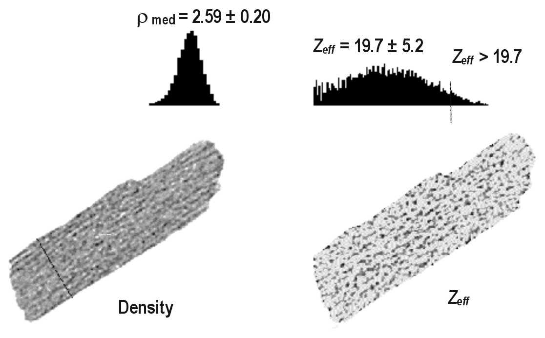
Identifying of minerals' abundance by Gamma- ray tomodensitometry
Paper Abstract A fragment of a polisulfide rock containing quartz and high-density mineral inclusions has been investigated by using a 192 Ir gamma-ray dual computer tomograph (Rizescu, C., Georgescu, Gh, Duliu, O. (2001) High density mineral inclusions in rocks evidenced by Gamma-ray tomodensitometry, Journal of Trace and Microprobe Techniques, Volume 19, No. 1, 119 –129). The tomographic image reveled the existence of three different mineral fraction, whose densities and effective atomic numbers were equal to 2.57 ± 0.24 g/cm3 (Zeff = 11.6 ± 2.1), 4.01 ± 0.22 g/cm3 (Zeff = 25.1 ± 2.1) and 6.01 ± 0.90 g/cm3 (Zeff = 68.5 ± 6.0) respectively. For this complex polymetallic rock we have identified the existence of three different mineral fractions: light (quartz), medium (mainly pyrite, blenda, chalkopyrite, and sphalerite), and heavy (galena). More, we have been calculated the abundance of each fraction for the entire sample: 70.6% for quartz, 26.8% for medium fraction and 2.6% for galena. A detailed mineralogical analysis confirmed these data. go to top
INVESTIGATION OF GEOLOGICAL SAMPLES
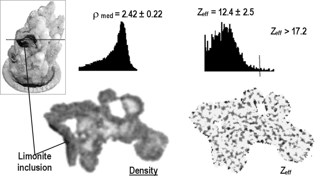
Paper Abstract Three specimens of metamorphic and sedimentary rocks were investigated using 192-Iridium dual energy gamma-ray computer tomography. (Duliu, O., G., Rizescu, C., Rickman, C., (2003) Dual energy gamma-ray axial computer tomography investigation of some metamorphic and sedimentary rocks, Neues Jahrbuch für Geologie und Paläontologie Abhandlungen, 228 (3), 343-362). Using this methodology, tomographs, depicting density as well as effective atomic number distribution with a 0.3 mm resolution, were obtained. Various details were evidenced including the granular structure of augen gneiss, the well-developed foliation pattern of mica rich gneiss, the banded configuration of nummulitic limestone or the concentric disposition of calcite microcrystals in a speleothem. In addition, by using the corresponding image histograms, it was possible to identify both major and minor components of investigated rocks. go to top
Nummulitic limestone
CT images (density and Zeff) indicate a layered disposition of individual nummulites. The investigated rock is composed by a great number of nummulites, disposed more or less parallel, and bound together by a cementing materiel which contains heavy fractions as iron or manganese minerals.Quartz-feldspar shale
The investigated section consists of 13 layers with an average thickness of 1.54 mm. A lighter material, depicted in more pale hues separates each lamina, which appears in darker tones. The investigated structure consists of an alternation of quartz and feldspar layers separated by muscovite rich strata.Hydrothermal quartz
The lighter components are quartz with a porous structure, i.e. micro-crystals aggregation with linear sizes smaller than 0.3 mm. On the left side of density CT image appears a thin layer of a heavier compact material, formed by most probably by limonite. go to top
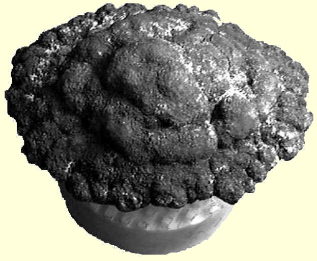
3-D investigation of polymetallic nodules
Paper Abstract Polymetallic nodules collected from the north-east Pacific Clarion-Clipperton region were investigated with 192-iridium dual-energy gamma-ray computer tomograph (Rizescu, C., Georgescu, Gh, Duliu, O., Szobotka, S., (2001) 3-D dual gamma-ray computer axial tomography investigation of polymetallic nodules, Deep Sea Research, Part I: Oceanographic Research Papers, Volume 48, 2529 – 2540).
Sixty-five successive digital sections, each with a thickness of 0.8 mm, reveal the 3-D density as well as the effective atomic number distribution function over the entire nodule volume, allowing one to obtain virtual sections across any desired plane. The tomograms show many details of internal structure of the investigated nodule such as and old nucleus fissure networks or layer structures. The total concentration of iron and manganese that determined by this method was equal to 19.4 ± 1.7. go to top
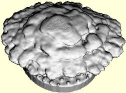
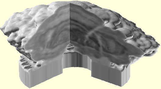
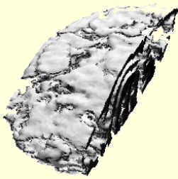
3-D image of nodule material whose density is greater than 1.91 g/cm3 Click here to see 3-D animation
Sedimentary core collected from the south-east Black Sea (near Bosphorus region at 1.400 m depth) were investigated with 192-iridium dual-energy gamma-ray computer tomograph.
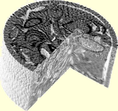
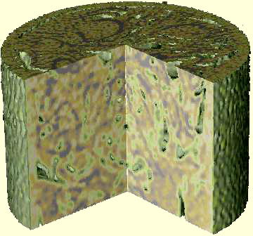
It can see disturbed intervals, sedimentary structures, lithologic heterogeneity, fracture orientation, permeability barriers etc. Scanning can reveal fractured systems, lithologic identification and can determine bulk density, porosity, sand/shale rations, saturation etc. go to top
Paper Abstract A comparative investigation of the same specimen of scleractinian Fungia fungites L. coral skeleton performed by means of both X- and Gamma-ray computer axial tomography has revealed several features such as annual growyh rings, gastrovascular cavity or scleorosepta to sixths order and more (Duliu, Gh., O., Preoteasa, F., Rizescu, C., Georgescu, G., (2004) X and Gamma - Ray CAT Investigation of Fungia fungites L. (Cnidaria, Scleractinia) Coral Skeleton, Journal of Trace and Microprobe Techniques, Volume 21, No. 3, 493 - 500). The apparent thickness of scleorosepta varied bewtween 0.1 and 0.19 mm while the number of growth rings allows us to estimate the average annual growth ratio to about 8 mm/year. Although X-ray computer axial tomography images appear to be of a better quality, no significant differences concerning sensitivity or spatial resolution could be noticed between the two categories of tomographs. go to top
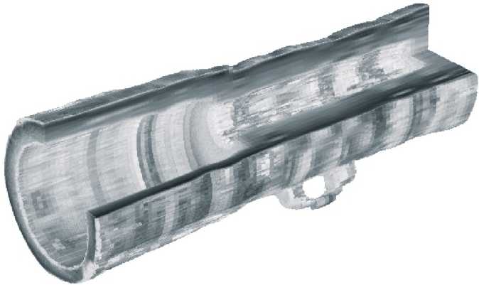
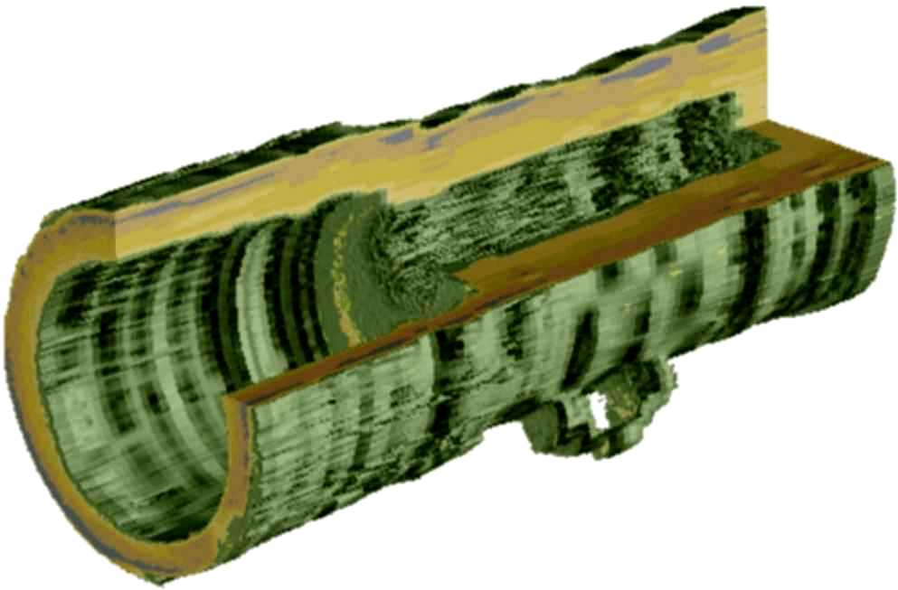
Complex Analysis of the "Cannon of Giurgiu" by Computerized Tomography with 192Ir
Paper Abstract The historical piece of artillery 'Cannon of Giurgiu' is investigated by computed tomography and X-ray diffraction methods (Rizescu, C., Beşliu, C., Jipa, A., Neacşu, I., (2003) Complex analysis of the “Cannon of Giurgiu” by Computerized Tomography with 192Ir, Journal of Archaeological Science, Volume 29, No.3, 267 – 275). 30 cross sections transversal to the cannon axis, spaced at 5 to 20 mm, are analyzed by dual-energy CT method with 192Ir radioisotope. Samples of corrosion material from the cannon pipe are tested by X-ray diffraction method. Numerous casting defects, large internal defects grown during cannon use, as well as severe corrosion regions have been detected. Structural analysis provides estimations of local material density and effective atomic number, leading to the identification of three domains in the cannon internal structure (bronze basic material, intermediate material and corrosion material). The degrading process is deep, with thickness of the corrosion layer ranging between 3.5 mm and 10 mm. These results determine the choice of appropriate restoration and conservation procedures.
Photos of "Cannon of Giurgiu"
The 'Cannon of Giurgiu' was discovered in 1981 at the medieval fortress from Giurgiu (city on the left bank of Danube river, 60 km S-SE of Bucharest). The 'Cannon of Giurgiu' is known as the oldest artillery object in Romania, dated as belonging to the period of Mircea the Old - prince of Wallachia between 1386-1418. It is 405 mm long, has a maximum diameter (at the end of the cannon pipe) of 147 mm, and weights about 23 kg. Other features are:
- the cannon head has 120 mm mean diameter and 42 mm length;
- the fire chamber has the diameter between 34 mm (at the beginning) and 40 mm (at the end), and a length of 168 mm;
- the cannon pipe has the interior diameter varying from 86 mm (at the beginning) and 106 mm (at the end), and the length of 195 mm;
- the cannon handle has a total length of 70 mm, and the diameter of its orifice is about 14 mm.
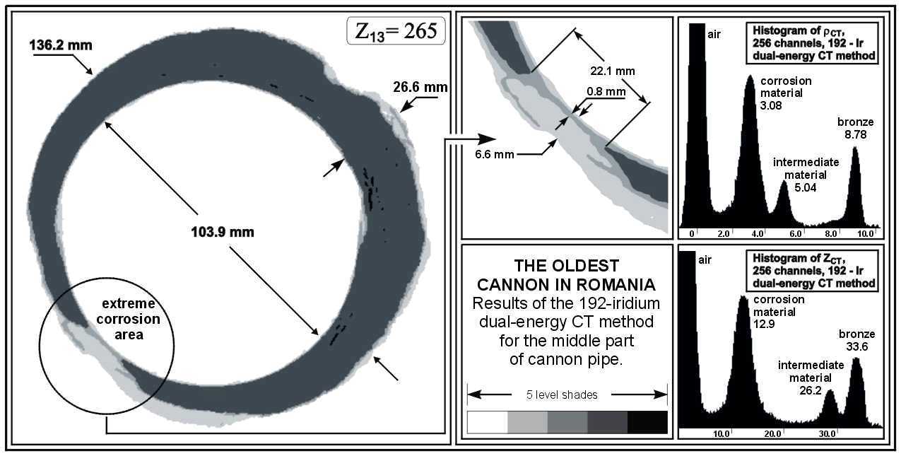
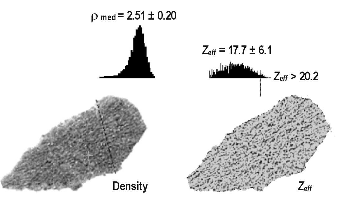
(Left) The results of the dual-energy CT method with gamma-ray 192-Iridium radioisotope for the extreme corrosion zone of the cannon pipe. The density distribution map of the investigated slice is presented on the left side of the figure. In the central part, it shows the effective atomic number distribution map for the region of interest. The histograms of density and effective atomic number in the investigated region are presented on the right side of above figure.
Analysis of material parameters determined by dual-energy CT and X-ray diffraction methods provides the following important information:
- The effective atomic number (Zeff = 33.6) and density (density= 8.78 g/cm3) of bronze correspond to the following material composition: 91.4%Cu7.8%Sn0.8%Pb. This formula is typical for bronze materials cast in the Middle Age metallurgic workshops.
- The intermediate material with Zeff = 26.2 and density= 5.04 g/cm3 corresponds to a structure of the following type: 45% basic material (bronze) and 55% corrosion material.
CURRICULUM VITAE PUBLICATIONS HOME Manufacturing & Technology Contact Information
| Copyright 2004, All rights reserved |