 |
|
|
|
|
|
|
|
|
|
|
|
|
|
|
|
|
|
|
|
|
Interesting intraoperative findings |
|
|
|
 |
|
|
|
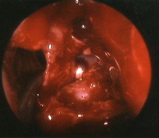
The picture above demonstrates the natural drainage pathway for the bulla ethmoidalis: the majority of the anterior ethmoid cell system. It typically drains posteriorly into the retro-bullar space. The probe is seen passing into the retro-bullar space and the tip is seen through the drainage pathway. This is on the patient's left side. These are very normal findings, but they are not often visualized this well, or discussed in textbooks. |
|
|
|
 |
|
|
|
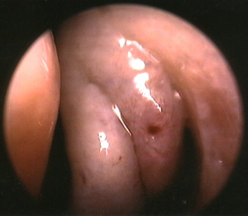
The picture above demonstrates an interesting finding. It looks as though the patient has two middle turbinates on her left side. Actually the more lateral "middle turbinate" is an air cell connected fully with the agger nasi cell above. It seemed to be an inferior extension of the agger nasi, or a concha bullosa that "pinched off" to form a psudo-turbinate. The uncinate process remained in place lateral to the turbinate, so it was not a pneumatization of the uncinate. |
|
|
|
On to more interesting photos2 |
|
|
|
On to more interesting photos3 |
|
|
|
On to more interesting photos4 |
|
|
|
Return to start |
|