|
||||||
|
Illusions and Paradoxes Seeing is Believing? - external link Jessica's visual experience. - life with Jessica. Jessica's Visual Fields - picture. Jessica's eyesight as the result of the Tumour. Jessica's expected eye surgery.
Visual Perception - See
also ---The Joy
of Visual Perception: A Web Book Very briefly, it may seem a silly question to ask what are our eyes for? However being able to see images or detect light is only part of what we do with our eyes. Seeing is one thing, knowing that we see and what we see is another. The biggest issue Jessica, and others like her, has is that her eyes do not see images very well anymore, however she is aware of the meaning of the world around her. Unless our brain knows what to do with the visual input then we may as well be blind! Our eyes are linked straight to our brains so that their sensory input goes directly into our brain where it is processed. Unless our brains are trained from an early age to correctly interpret the world our eyes reflect then none of us will know the meaning of the world around us. Seeing and knowing what we see and its meaning are part of a complex system called Visual perception or Cognitive perception. Though we have 5 senses it is our sense of sight which seems to do the most to assist us interpret our world - or so we think while we can see. When someone losses any one of their 5 senses those left become more acute to compensate for the loss. When people lose their sight their other 4 senses take up some of the slack, so to speak, to make up for their lack of vision. This explains why Jessica has extremely good mind maps of the City of Sydney and other places. Vision impaired people make the best use of touch, taste, smell and sound to compensate for their loss of vision or partial loss thereof. Think about it! If someone was told as a child that the colour green is called blue then what colour are the trees? Life with Jessica Visual experience. I have a wonderful, marvellous wife who lives a full life under extremely difficult circumstances. Jessica lost her sight as the result of the Pituitary adenoma - a Prolactinoma to be exact. The tumour, situated on the posterior lobe of the Pituitary gland, grew so that it pressed upon the Optic chiasm and eventually brought about the loss of her sight. Jessica still has some sight in her left eye, t hough now experiences the condition known as Bilateral Hemianopia. This means that her small percentage of sight is missing its outer visual field due to damage to the Nasal nerve. Below you will read part of an article from Wikipedia about the Optic chiasm, it will assist you if you make your way to the site to read the definition there. When Jessica and I first met her loss of vision wasn't immediately obvious. She did not even use her sight stick so as not to draw attention to herself, however I had not known her for very long before I realised that she needed to use the sight stick to prevent herself bumping right up against people and to indicate that there was a problem with her vision. Over the eight years we have been together her sight has become progressively worse until now she sees with difficulty when she is out and requires the use of a cane with a roller ball to navigate her way around the City and outer suburbs. Due to her frequent seizures it is more often that not necessary for her to be guide by myself or one of the day cares who come to relieve me. Nevertheless, Jessica makes the maximum use of her remaining vision, she can often find things on the supermarket shelf quicker than I since she knows how to use her bit of vision to scan the shelves progressively until she finds whatever we are looking for. In the time we have been together we have put a few computers together, Jessica still works from the command prompt and does the maintenance on our computer. She also enjoys her digital camera and takes excellent photos just by pointing and shooting. She will then work on the photos in our imaging programs, Micrografx and Image Pro 10, to bring them up to scratch and to make Panoramas and stitches which reveal entire vistas such as the coastline in Kiama near to the Blowhole caravan park. You will see some of her pictures once we load the galleries. It is amazing what people can do to compensate for the loss of one of their senses, sometimes I might believe that my wonderful wife is not visually impaired especially when I see her wielding the electric drill to repair or create something. She cuts my hair and uses the sewing machine and she loves to wash which is not so good since I am t he one who has to do the ironing.
<- Click to enlarge.
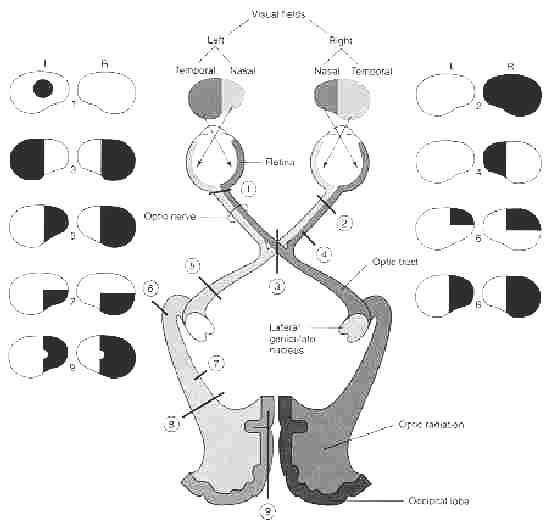
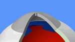
Knowledge
of the topographical arrangement of visual fibres as they travel in the chiasm is of utmost importance to understand the effects of compressive lesions in this
portion of the optic pathway. The optic nerves from both eyes meet and cross at the optic chiasm, at the base of the frontal lobe of the brain. At this point the information from both eyes is combined and split according to the field of view. The corresponding halves of the field of view (right and left) are sent to the left and right halves of the brain, respectively (the brain is cross-wired), to be processed. That is, though we might expect the right brain to be responsible for the image from the left eye, and the left brain for the image from the right eye, in fact, the right brain deals with the left half of the field of view, and similarly for the left brain. (Note that the right eye actually perceives part of the left field of view, and vice versa). Jessica's eyesight as the result of the Tumour.
Bitemporal hemianopia is a specific type of visual disturbance in which
sight in the outer half of the visual field of each eye is lost. As a
result, the patient retains central vision but loses sight at the edges of
his or her vision. This is not always obvious to him or her, even when the
damage is quite severe on objective testing, because one tends to focus
attention more on objects in the centre of the visual field. Jessica's Visual Fields - picture.
How does it affect daily living?
Hemianopia can cause loss of confidence with doing
simple daily tasks. For example: The person may feel unsafe crossing the
road or going shopping, Loss of half the visual field will affect many
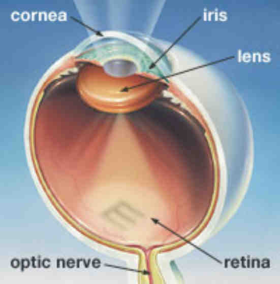
daily tasks including reading, travel and daily living skills. This photo with thanks from Special Kids in the UK In Jessica's case she has her near blindness in her favour, also the experience of having had the condition for some time. In the case of those who acquired the condition recently it will take time for them to reinterpret the world they see and to train themselves to fill in the missing parts of their visual field input. The diagram below shows how loss of vision varies depending upon at which level in the visual l system is damaged, see below for ledgend. See 3 of this diagram. Follow the link to the web for a clearer picture.
· 1
= central scotoma secondary to optic neuritis (does not respect the
vertical meridian)
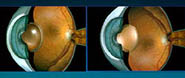
Definition-Simply put, a cataract is a "clouding" of the lens in your eye. The lens, located just behind the iris, or the coloured part of your eye, works like the lens of a camera. It picks up images, then focuses the lights, colours, and shapes on the retina - the transmitter located at the back of your eye that sends the images to your brain. Normal lens v cloudy lens What is the cornea?
Corneal transplantation "What is corneal transplantation? A corneal transplant is the way of removing your damaged cornea and replacing it with a healthy cornea from the eye of a suitable donor. The donor will be a person who has (or whose family has) given consent for their corneas to be used for medical purposes after their death. The donor cornea is thoroughly checked and prepared to ensure there is no possibility of it being infected.
Please read their web page
for further information about Corneal Transplantation. Also seeKeratoconus Australia - Resources Everything you wanted to know about corneal transplants and their care. ... Types of contact lenses for keratoconus ... Corneal Donation Service, corneal donation and corneal grafts in Australia ...
Aftercare - care of Laser and Corneal surgery Associates.P.C. also ienhance.com N.B. It is very difficult to find clear information regarding the aftercare of the graft. snip
"RETURN TO ACTIVITY
|
||||||